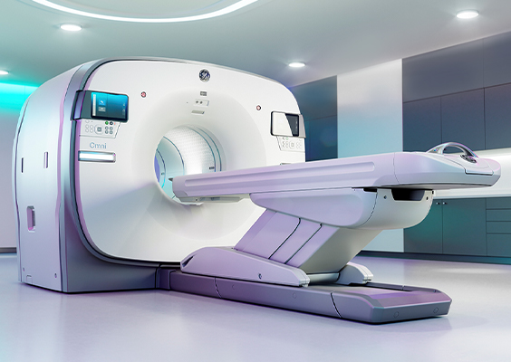
PET-CT
What is a PET-CT?
PET-CT is a cutting-edge diagnostic device that combines positron emission tomography (PET) and computed tomography (CT).
PET is used to determine the spread of tumors, status of metastasis, and effectiveness of treatment, and to diagnose recurrences by injecting fluorodeoxyglucose (FDG), a glucose metabolism indicator, and monitoring the status of glucose metabolism in the body.
By simultaneously imaging with CT, it is possible to precisely locate lesions detected by PET. It allows evaluating almost the entire body within a scanning time of approximately 10-15 minutes.
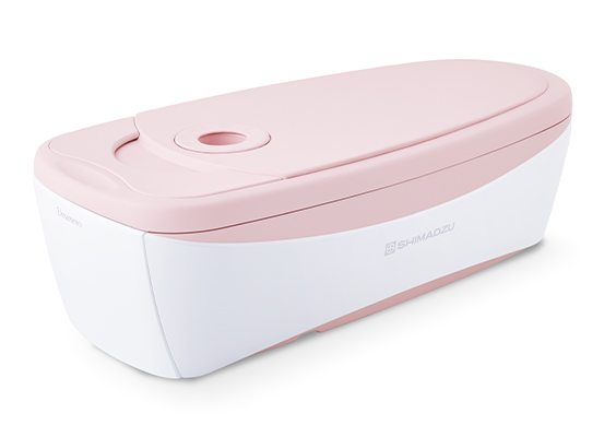
What is a Breast PET?
This is a diagnostic test using a breast-specific PET device capable of detecting small breast cancers that are difficult to find. Patients lie face down on a special bed, with their breast placed in the detector hole, which allows testing without any pressure on the breast. The test is not significantly affected by “high-density breasts”, and can be performed even after breast augmentation surgery because it does not involve pinching the breast.
- This test must be conducted in combination with a whole-body PET-CT scan.
- It is conducted consecutively with a whole-body PET-CT scan; therefore, the FDG drug is administered only once, and no additional administration is required.
Issues to consider prior to a PET-CT
Medical considerations prior to the PET-CT may includes;
- Diabetes
If you are under treatment for diabetes, please consult with your doctor in advance regarding medication and insulin injections. - Pregnancy
As a general rule, we do not perform the test if you are pregnant or have the possibility of pregnancy. Please inform us in advance. - Exercise
Please refrain from any strenuous work or exercise before the examination.
PET-CT procedure
Duration approx. 120 min
-
STEP 1
Medical interview -
STEP 2
After measuring blood glucose level, 18F-FDG is injected intravenously. -
STEP 3
Rest in a resting room for about 60-75 minutes. -
STEP 4
Urinate before the examination. -
STEP 5
Scanning for about 10-15 minutes.- Breast PET: If you are undergoing a breast PET scan, scanning will be performed for another 15 minutes after the whole body scanning.
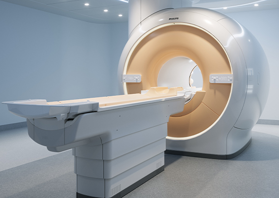
MRI
What is a MRI scan?
This is a diagnostic test in which patients are placed inside a tunnel with a strong magnetic field to image the inside of their body using the power of magnets and radio waves.
It allows visualizing cross-sections of the body from various angles, providing detailed information on the brain, spinal cord, internal organs, joints, and blood vessels.
In addition, as it does not use X-rays, there is no radiation exposure.
Issues to consider prior to a MRI
Because MRI scans use strong magnets and radio waves, great care must be taken to prevent accidents and burns.
Medical considerations prior to the MRI scan may includes;
- Metal
- Pacemaker
- Implantable cardiac defibrillator
- Vascular clips or wires
- Cochlear implant
- Intracranial pressure valve
- Artificial blood vessels
- Artificial heart valve
- Stent installed less than 2 months
- Continuous Glucose Monitoring tool
- Dentures or magnetic dentures
- Transdermal skin patches/nicotine paches
- Contraceptive ring
- Tattoo / Tattooed makeup
- Tattoos and permanent makeup may cause burns or discoloration.
- Pregnancy
As a general rule, we do not perform the test if you are pregnant or have the possibility of pregnancy. Please inform us in advance. - Claustrophobia
Since the examination involves entering a narrow tunnel, people with a tendency toward claustrophobia may feel more anxious. Please relax as much as possible during the examination.
MRI scan procedure
Duration approx. 15 min / per region
-
STEP 1
Medical interview -
STEP 2
Scanning- Please hold your position during the examination.
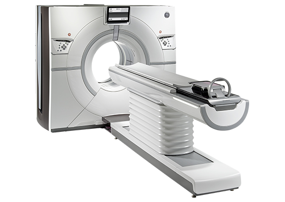
CT
What is a CT scan?
In a CT scan, X-rays are irradiated from around the body, and the X-rays passed through the body are processed by a computer to obtain cross-sectional images (slices) of the body. It allows visualizing the internal structure of any part of the body and provides detailed information on the inside of the body that cannot be obtained with regular X-rays.
- CT scans do involve radiation exposure, as they use X-rays. We make every effort to keep the exposure level to a minimum.
Issues to consider prior to a CT
Medical considerations prior to the CT scan may includes;
- Pregnancy
As a general rule, we do not perform the test if you are pregnant or have the possibility of pregnancy. Please inform us in advance. - Barium
CT scan cannot be performed if the patient has had a barium test within 5 days prior to the test.
CT scan procedure
Duration approx. 10 min / per region
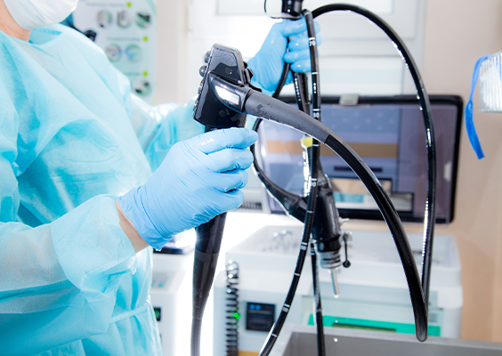
Upper endoscopy
What is an upper endoscopy?
Upper endoscopy is a medical examination that uses an endoscope to observe the esophagus, stomach, and duodenum.
About Nasal Endoscopy
- If you wish to undergo a nasal endoscopy, please inform us in advance.
- Those with narrow nasal passages or a deviated septum may not be suitable candidates for nasal insertion. In cases where nasal insertion is difficult in both nostrils, you may need oral upper endoscopy.
- After the examination, you may have nasal discharge, nosebleeds, or nasal discomfort.
- Those who are breastfeeding are not eligible for this examination.
Attention
- If you can not tolerate the procedure, there will be no refund.
- If you cancel the upper endoscopy on the examination day, rescheduling for another date will not be possible.
About Sedation
Only available at the facilities in Kyoto
- You have the option to undergo the examination with the use of sedatives, which can alleviate anxiety and discomfort during the examination.
- Sedatives do not guarantee complete unconsciousness.
- After the examination, you will be observed for a period of time, while the sedative medication weans off.
- Please be aware that individual responses to sedatives may vary, and they may not be effective in some cases.
- There is an additional fee of 12,100 yen for the use of sedatives.
Facilities offering Sedation
- Oike Clinic
- Oike Clinic Ladies Plaza
The following are not eligible for Sedation
- Those aged 72 and above
- Lactating mothers
- Those who have glaucoma
- Those who have myasthenia gravis
On the day of the procedure, please abstain from
- Driving a car or motorcycle.
- Wearing nail polish to allow for easier monitoring of your oxygen level from your fingertips.
Upper endoscopy procedure
Duration approx. 10 min
-
STEP 1
You will ingest medication to clean the mucous membrane of the stomach.- For nasal endoscopy, a topical decongestant will be sprayed into the nasal cavity to prevent nosebleeds and let the nasal endoscope pass easily through your nasal cavity.
-
STEP 2
A gel type anesthetic will be injected into your throat.- For nasal endoscopy, an anesthetic will be injected into the nasal cavity.
-
STEP 3
An endoscope will be inserted through your mouth or nose to observe the esophagus, stomach, and duodenum. -
STEP 4
If sedation is administered, you will need to take a period of rest until the sedatives wear off.